
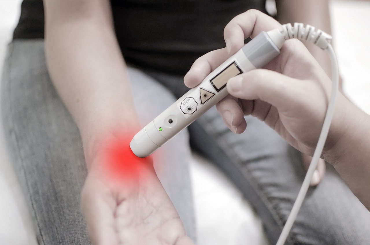
Rheumatoid arthritis is a disease characterized by chronic inflammation of synovial tissue in joints, potentially leading to permanent damage to cartilage and bone. The chronic nature of this inflammation can lead to a range of symptoms, including pain, stiffness, and swelling in the affected joints, which can significantly impact a patient’s quality of life. If left untreated or inadequately managed, rheumatoid arthritis can result in permanent damage to the joints, affecting physical function and overall well-being.
Researchers have found that PET/CT imaging can predict treatment response in patients with rheumatoid arthritis, allowing for earlier determination of which patients are most likely to respond to treatment. This is particularly important, as the current standard for determining treatment efficacy can take three to six months, during which time patients may continue to experience symptoms and potentially suffer from further joint damage. By utilizing PET/CT imaging, healthcare providers may be able to identify non-responders to treatment earlier, enabling them to pursue alternative therapies that may be more effective.
According to the report, this new imaging technique could help determine the patients who are most likely to respond sooner, allowing non-responders to pursue more effective therapies. The ability to predict treatment response early on can significantly improve patient outcomes, as it enables healthcare providers to tailor treatment plans to individual patients’ needs. This personalized approach to treatment can help minimize the risk of unnecessary side effects, reduce healthcare costs, and improve overall quality of life for patients with rheumatoid arthritis.
The current standard for determining treatment efficacy can take three to six months. During this time, patients may be required to undergo regular assessments and monitoring to evaluate the effectiveness of their treatment. However, this prolonged period of uncertainty can be challenging for patients, who may experience ongoing symptoms and anxiety about their treatment outcome. The use of PET/CT imaging has the potential to significantly reduce this waiting period, enabling healthcare providers to make more informed decisions about treatment earlier on.
Anti tumor necrosis factor is a successful treatment for many patients with rheumatoid arthritis; however, it is only effective in 50% to 70%. This variability in treatment response highlights the need for more effective tools to predict which patients are most likely to benefit from this therapy. By identifying non-responders earlier, healthcare providers can explore alternative treatment options that may be more effective, reducing the risk of disease progression and improving patient outcomes.
PET/CT Imaging Technique
A new PET/CT imaging technique has been utilized to quantify macrophages and determine their association with clinical disease activity. Macrophages, a type of white blood cell, play a key role in the development and continuation of rheumatoid arthritis, and are a promising biomarker for assessment and monitoring of disease activity. The use of PET/CT imaging to quantify macrophages offers a non-invasive and objective method for evaluating disease activity, which can be used to inform treatment decisions and monitor patient response to therapy.
Wouter van Binsbergen, MSc, MD, PhD, a student at the Amsterdam University Medical Center, said that “In our study, we utilized a new PET/CT imaging technique to quantify macrophages and determine their association with clinical disease activity.” This study demonstrates the potential of PET/CT imaging to provide valuable insights into the underlying biology of rheumatoid arthritis, enabling healthcare providers to develop more effective treatment strategies.
In the study, researchers conducted whole-body 11C-DPA-713 PET/CT scans on 20 rheumatoid arthritis patients undergoing aTNF treatment. The use of whole-body scanning enables researchers to evaluate disease activity across multiple joints, providing a full understanding of the patient’s condition. By quantifying macrophages in this way, researchers can gain a better understanding of the underlying mechanisms driving disease activity, which can inform the development of more effective treatments.
Study Findings
Quantitative 11C-DPA-713 PET/CT measurements at baseline and four weeks after aTNF treatment showed significant association with clinical disease activity at 26 weeks. This suggests that PET/CT imaging can provide an early indication of treatment response, enabling healthcare providers to make informed decisions about ongoing treatment. The ability to predict treatment response at an early stage can significantly improve patient outcomes, as it enables healthcare providers to adjust treatment plans accordingly.
They found that the addition of specific clinical data to SUV data of certain joints improved the potential predictive value in multivariable regression analyses at baseline and 4 weeks in specific instances. This highlights the importance of combining imaging data with clinical information to gain a more full understanding of disease activity. By integrating these different data sources, healthcare providers can develop a more subtle understanding of patient response to treatment, enabling them to make more informed decisions about ongoing care.
These findings show that non-invasive assessment and monitoring of macrophages using PET/CT has value to predict very early potentially already at baseline the outcome of anti-TNF treatment in rheumatoid arthritis patients. The use of PET/CT imaging in this way offers a promising approach to personalized medicine, enabling healthcare providers to tailor treatment plans to individual patients’ needs. By predicting treatment response at an early stage, healthcare providers can minimize the risk of unnecessary side effects, reduce healthcare costs, and improve overall quality of life for patients with rheumatoid arthritis.
Clinical Application
Related: European Radiology Unites at ESOR’s 20th Anniversary